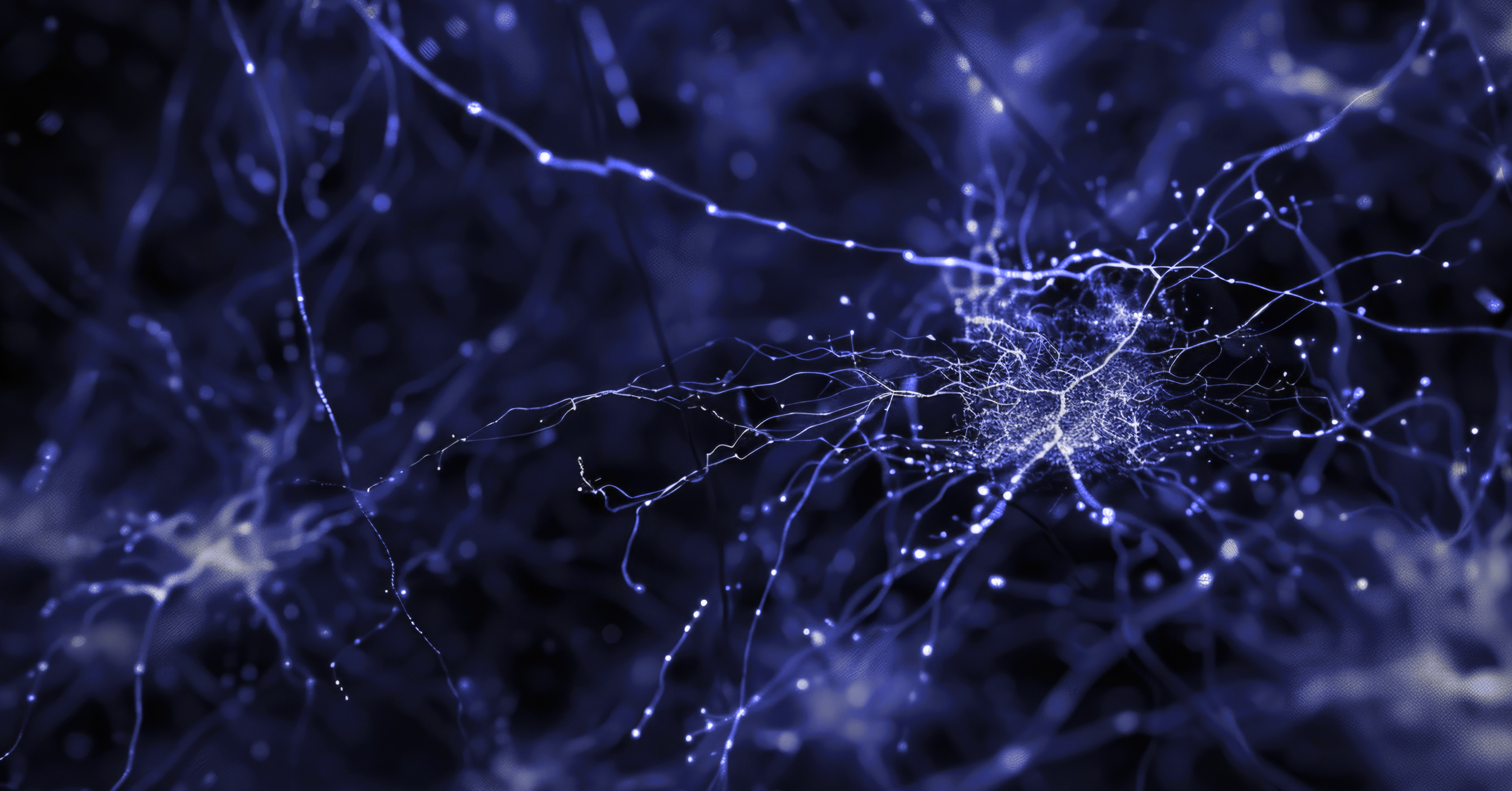
•  exploring the science

A New Way to Measure Brain Health

Resonant currently offers a research-use only (RUO) platform that enables cell-type-specific profiling of neuron- and glia-derived cfDNA from human blood plasma. 

By quantifying signals from key brain cell populations, our assay offers a minimally invasive approach to studying neurodegeneration, powering discovery, biomarker development, and therapeutic research across Alzheimer's, Parkinson's, ALS, and related conditions.

Contact us

our approach

•  exploring the science

Cell-Type Resolution
Across the CNS

This multi-lineage resolution allows researchers to investigate region- and cell-type–specific signatures of neurodegeneration, providing a dynamic view of cellular turnover in the central nervous system (CNS).

Resonant’s assay quantifies cfDNA originating from six key neural and glial populations:

Cortical neurons

Dopaminergic neurons

Spinal motor neurons

Astrocytes

Microglia

Schwann cells

Deciphering the Data

Predicting Cell Fractions At Cell Specific DMR's

Figure 1. Primary cell-of-origin classifiers demonstrate strong linearity when applied to in silico cfDNA-like admixtures for neurons and glia. Classifiers for primary cortical (A, R² = 0.998), dopaminergic (B, R² = 0.993), and spinal motor (C, R² = 0.876) neurons, as well as astrocytes (D, R² = 0.993), Schwann cells (E, R² = 0.986), and microglia (F, R² = 0.992), showed high concordance between predicted and actual DNA fractions across biologically relevant concentrations. Read length distributions are shown in G–H. gDNA reads were computationally fragmented to mimic native cfDNA profiles and better reflect biological signal. 

Linearity Across Inputs

To evaluate platform performance at various concentrations, we created in silico admixtures, mixing fragmented neuronal and glial gDNA reads into blood plasma reads (0%, 0.1%, 1%, 10%, 15%, 25%, 50%, 75%, and 100%). 

The classifiers demonstrated strong linearity across all six primary cell types, suggesting cfDNA signals scale proportionally with biological input rather than assay noise. This supports their potential utility for tracking cell type-specific degeneration, monitoring therapeutic responses, and assessing disease dynamics over time.

•  exploring the science

Validating The Biomarker

1. Clinically Sourced Samples

We analyzed cfDNA from plasma samples collected from controls and patients with Alzheimer’s, Parkinson’s, ALS. These clinical samples enabled real-world assessment of across neurodegenerative conditions.

2. Multi-Cell-Type Modeling

We developed multivariate models that integrate cfDNA signals from six CNS cell types. This approach outperformed single-marker strategies, capturing coordinated, disease-specific patterns of cellular injury and turnover.

3. Accurate Disease Classification

Our integrated models enabled >98% classification accuracy across AD, PD, and ALS cohorts. Leveraging the full spectrum of cell-type inputs, the platform distinguished disease presence and type in research-use settings.

Explaining The science‍

Classifying Neurodegenerative Disease with Cell-Type–Specific Molecular Signatures

Individual neuron classifiers detected elevated cortical, dopaminergic, and spinal motor neuron-derived cfDNA in the plasma of patients with Alzheimer’s disease, Parkinson’s disease, and ALS, respectively, reflecting cell type-specific neurodegeneration. Integrating these three neuronal signatures into a single multivariate cell-of-origin model further improved disease detection accuracy, outperforming both individual neuron classifiers and iPSC-derived models across all disease contexts.

Download pdf

Figure 2. Combined primary neuron classifiers outperform individual neuron and iPSC-derived models. A multi-neuron classifier integrating cortical, dopaminergic, and spinal motor signatures achieved near-perfect accuracy in detecting Alzheimer’s (A, n = 35, AUC = 1.000, 95% CI: 1.000–1.000), Parkinson’s (B, n = 45, AUC = 0.985, 95% CI: 0.969–1.000), and ALS (C, n = 53, AUC = 0.991, 95% CI: 0.975–1.000). Performance exceeded that of individual neuron classifiers (D–F) and iPSC-derived models (G–I) in multi-disease contexts.

98%

Multi-Disease

Classification accuracy distinguishing between AD, PD, and ALS cases

99%

Parkinson's Disease

Classification Accuracy vs. Control

98%

ALS

Classification Accuracy vs. Control

99%

Alzheimer's Disease

Classification Accuracy vs. Control

•  exploring the science

Why Model Performance Across Disease Cohorts Matter

These findings suggest that cfDNA signatures derived from specific neural and glial populations reflect the cellular hallmarks of distinct neurodegenerative diseases. By modeling contributions from multiple neural and glial cell types, Resonant’s platform supports high-resolution detection of disease-relevant biology—which can be applied to both the identification and differentiation of neurodegenerative conditions. 

This capability provides researchers and clinical partners with a powerful molecular tool to study mechanisms of disease, stratify cohorts, and assess therapeutic effects through a non-invasive, cell-type–specific lens.

Resonant’s platform is for Research Use Only and is not intended for clinical diagnostic purposes. 

•  The Biology Behind the Biomarker

•  The Biology Behind the Biomarker

As brain cells degenerate, fragments of DNA are released into the circulation. This cell-free DNA (cfDNA) carries molecular features that reflect its cell of origin. By decoding those patterns, Resonant identifies which neuronal and glial populations are affected, and to what degree.

This approach links measurable cfDNA in blood to underlying neurodegenerative processes, enabling researchers to study disease biology with greater resolution and less invasiveness relative to conventional tools.

our solutions

Expanding Our View of Brain Biology

Enhanced Cell Subtype Identification

Expanded cell types, including cortical projection and hippocampal neurons, astrocytes, and microglia.

Multi-Modal Integration

Frameworks to integrate cfDNA signals with imaging, fluid biomarkers, and clinical phenotyping.

Increased Resolution

Improved detection of rare or low-abundance neuronal signals to capture early or subtle changes.

Translational Research Fit

Designed to support exploratory endpoints, from biomarker discovery to pharmacodynamic readouts.

our solutions

Translating Science Into Actionable Tools